


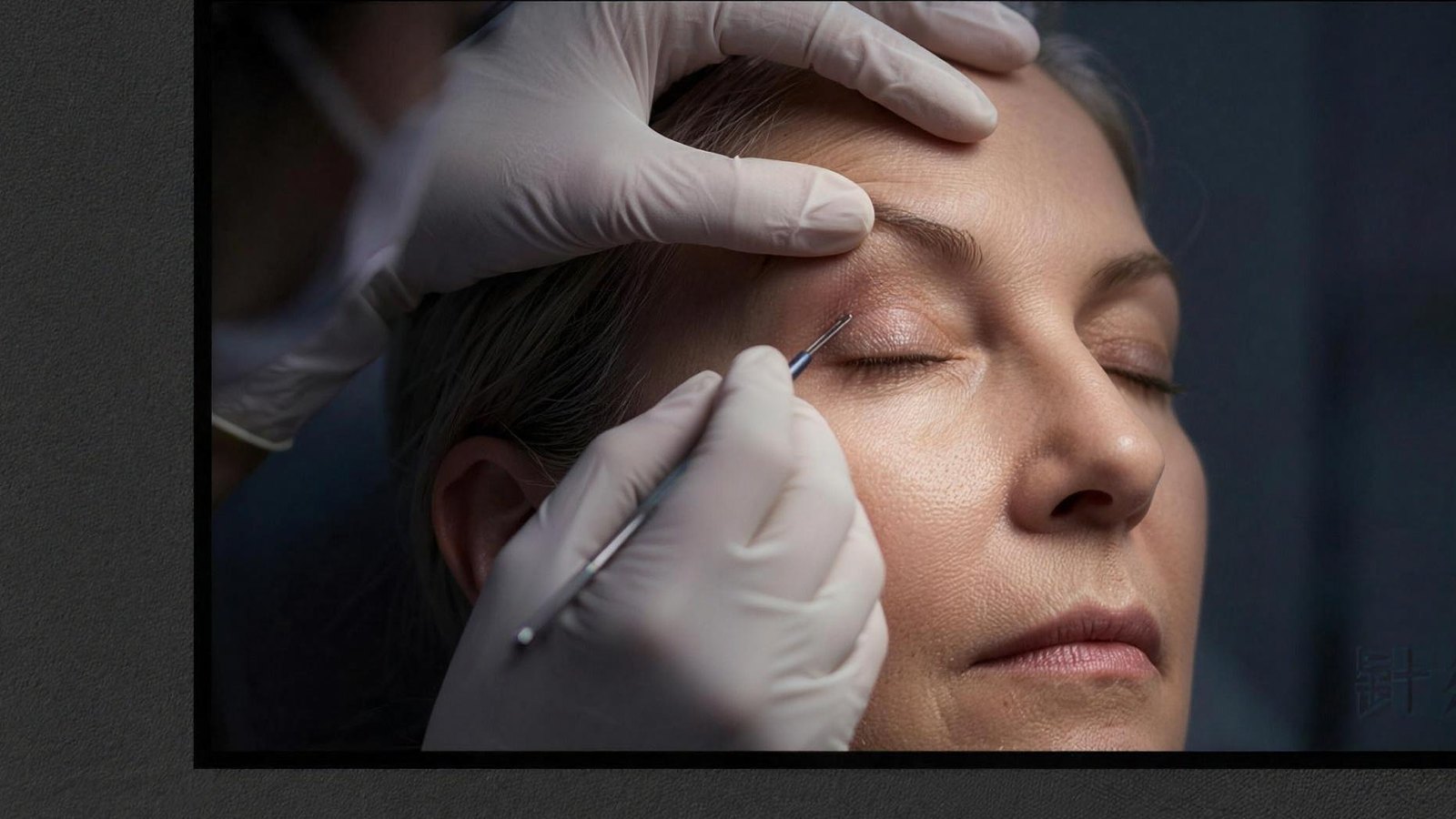
Eyelid ptosis may be present from birth, develop with age, or relate to nerve, muscle, trauma or previous surgery. Sudden drooping with double vision, a pupil change, weakness or severe headache needs urgent assessment. Planning depends on the cause, levator function, ocular surface and visual field. Surgery can result in under- or overcorrection, asymmetry, incomplete closure, dryness or a need for revision; a particular height or symmetry cannot be guaranteed. An examination appointment can be arranged at the Maslak clinic in Istanbul.
What Is Eyelid Ptosis?
Ptosis refers to the abnormal lowering of the upper eyelid margin. In a normal eye, the upper lid rests approximately 1-2 mm below the upper edge of the iris. When the eyelid drops further, it can partially or completely cover the pupil, interfering with vision. The severity of ptosis is measured by the margin reflex distance (MRD), which quantifies the relationship between the eyelid position and the center of the pupil.
Types of Ptosis
Ptosis is classified based on its onset and underlying cause. Congenital ptosis is present at birth and typically results from poor development of the levator muscle, which is responsible for lifting the eyelid. This type accounts for a significant portion of ptosis cases in children and may be associated with other developmental conditions.
Acquired ptosis develops later in life and encompasses several subtypes. Aponeurotic ptosis is the most common form in adults, resulting from stretching or dehiscence of the levator aponeurosis due to aging. Neurogenic ptosis arises from nerve damage or neurological conditions such as third cranial nerve palsy or Horner syndrome. Myogenic ptosis occurs in muscle disorders like myasthenia gravis. Mechanical ptosis results from eyelid masses, excess skin, or scarring that weighs down the lid. Additionally, traumatic ptosis can follow eye surgery or facial injury.
Recognizing the Symptoms
The signs of ptosis extend beyond the visible drooping of the eyelid. Patients frequently report reduced peripheral vision, difficulty reading, and eye fatigue. Many develop a compensatory habit of raising their eyebrows or tilting their head backward to see better. Furthermore, chronic brow elevation can lead to forehead tension and headaches. In children, untreated ptosis carries a risk of amblyopia (lazy eye), making early detection particularly important.
What Causes Eyelid Drooping?
Understanding the cause of ptosis is essential for determining the appropriate treatment approach. Age-related weakening of the levator aponeurosis accounts for the majority of adult cases. Over decades, repeated eyelid movements and gravitational forces gradually stretch the tendon that connects the levator muscle to the eyelid.
Neurological conditions represent another significant category. Third nerve palsy can cause sudden, severe ptosis along with eye movement abnormalities. Horner syndrome produces a mild ptosis accompanied by a smaller pupil on the affected side. Myasthenia gravis characteristically causes fluctuating ptosis that worsens with fatigue. Contact lens wear, particularly rigid lenses used over many years, has been identified as a contributing factor in some patients. Previous eye surgery, including cataract extraction and blepharoplasty, may also lead to ptosis in certain cases.
Eyelid Ptosis Treatment Options
The choice of eyelid ptosis treatment depends on the underlying cause, the degree of drooping, and the function of the levator muscle. Surgical intervention remains the primary approach for most cases, though the specific technique varies based on individual assessment.
Levator resection is the most widely performed procedure. During this surgery, the levator muscle or its aponeurosis is shortened to elevate the eyelid to its proper position. This technique works well when the levator muscle still has reasonable function. The procedure is typically performed under local anesthesia, and the surgeon can adjust the eyelid height during the operation.
Frontalis sling surgery is reserved for cases of severe ptosis with poor levator function, which is often the case in congenital ptosis. In this technique, the eyelid is connected to the frontalis (forehead) muscle using a sling material, allowing the patient to lift the eyelid by raising their brow. Muller muscle resection offers an alternative for mild ptosis cases and can be particularly effective when a positive response to phenylephrine testing is observed.
In some situations, ptosis repair is combined with other procedures. For instance, patients with both ptosis and excess upper eyelid skin may benefit from simultaneous blepharoplasty. Similarly, botulinum toxin applications may complement surgical treatment in select cases. The goal of any combined approach is to achieve both functional improvement and a natural, harmonious appearance.
Recovery Process After Ptosis Surgery
Recovery following eyelid ptosis treatment varies depending on the surgical technique used and the patient's overall health. Most procedures are performed on an outpatient basis under local anesthesia, allowing patients to return home the same day. Initial swelling and bruising around the eyes are normal and expected.
During the first week, cold compresses help reduce swelling. Sutures are typically removed within 5 to 7 days. Patients should avoid heavy lifting, bending, and strenuous exercise for approximately two weeks. Prescribed eye drops and ointments should be used as directed to prevent dryness and promote healing. Most patients can return to light daily activities within a week, though complete healing takes 4 to 6 weeks.
The final results of ptosis surgery may take several months to fully settle. Follow-up appointments are essential for monitoring healing progress and ensuring optimal outcomes. Patients are advised to wear sunglasses outdoors and avoid eye makeup during the initial recovery period. According to the American Academy of Ophthalmology, regular follow-up care plays a critical role in successful ptosis outcomes.
Risks and Considerations
As with any surgical procedure, ptosis repair carries certain risks. Under-correction, where the eyelid remains lower than intended, is one of the more common issues and may require revision surgery. Over-correction can lead to lagophthalmos, a condition where the eyelid cannot fully close, potentially causing corneal dryness and irritation. Asymmetry between the two eyelids is another possibility.
An important consideration in unilateral ptosis is Hering's law of equal innervation. When one drooping eyelid is corrected, the opposite eyelid may appear to drop, as the brain reduces the compensatory effort previously directed to both sides. Experienced surgeons account for this phenomenon during preoperative planning. Rare complications include infection, bleeding, and adverse reactions to anesthesia. Experience and careful planning may reduce risk, but they cannot eliminate asymmetry, under- or overcorrection, incomplete closure or revision.
Frequently asked questions
Why can sudden eyelid ptosis be an emergency?
New one-sided ptosis can occasionally reflect a neurological or muscle disorder. If it occurs with double vision, unequal pupils, restricted eye movement, facial or arm weakness, or a severe headache, do not wait for a routine appointment; seek urgent medical assessment to identify the cause.
How is ptosis distinguished from excess upper-lid skin?
In ptosis the eyelid margin sits low; with excess skin, a skin fold hangs over the lid. Brow descent can also alter the appearance. The clinician checks lid height, levator function, brow position, eye movements and ocular surface, adding visual-field or other tests when indicated.
Will ptosis surgery make both eyelids exactly equal?
The goal is to improve vision and eyelid position, but exact symmetry cannot be guaranteed. Height and contour may change during healing, and hidden droop in the opposite lid can become apparent. Undercorrection, overcorrection, dryness or incomplete closure may require monitoring, treatment or revision.
Related guides
Sources
- What is ptosis? — American Academy of Ophthalmology
- Ptosis Correction (Adults) — Cambridge University Hospitals NHS
This content provides general information, not a diagnosis or personal treatment recommendation. Suitability, risks and alternatives require an individual medical assessment.
