

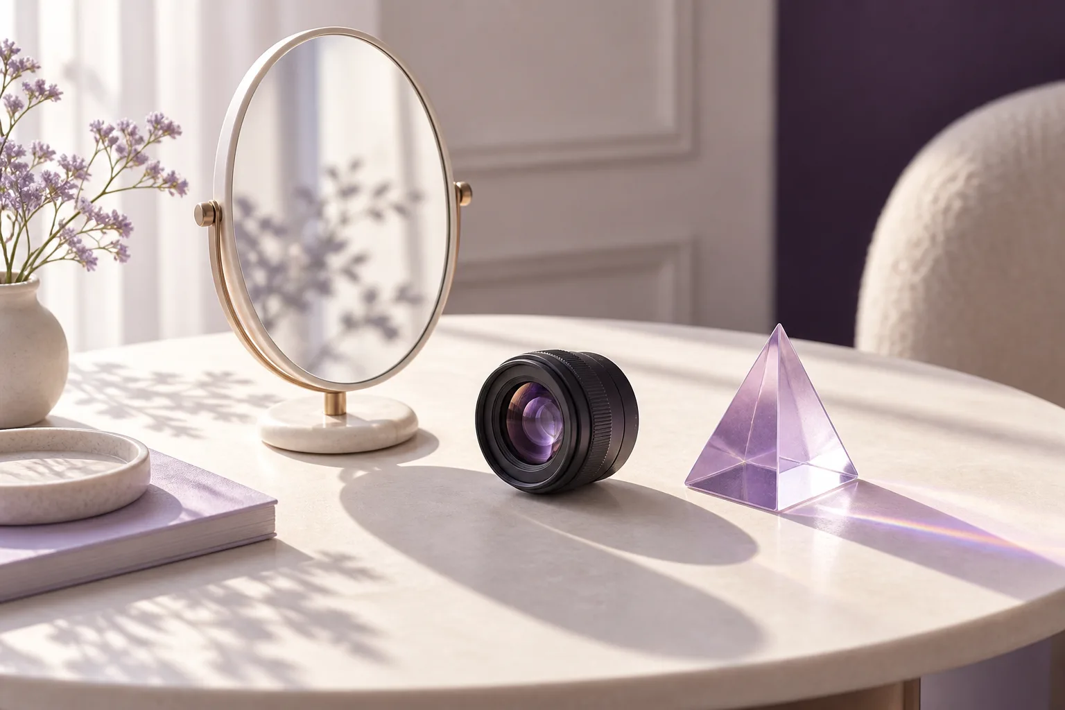
Your eye area may look familiar in the mirror yet appear more shadowed or less symmetrical in a photograph. Noticing this difference is understandable, but one image does not establish a new problem or a need for a procedure. A photograph records a particular distance, lighting setup and momentary expression. An examination also considers your symptoms and how your eyelids function.
The same face in different shooting conditions
Light from above can cast shadows beneath the contours around the eyes; more diffuse lighting changes how those shadows appear. Camera height and closeness to the face can also affect the proportions in an image. A bathroom mirror, a close selfie and a portrait taken farther away are therefore not equivalent comparisons. Consistent conditions are an important part of making clinical photographs meaningfully comparable.
This does not mean every difference between photographs is explained by lighting alone. A feature highlighted in one image may also be present in everyday life. Rather than trying to decide from a photograph exactly what comes from the camera and what comes from anatomy, describe the circumstances in which you notice it.
A mirror and a photograph are different views
In a mirror, you see yourself moving and can change your head position and gaze immediately. A photograph freezes just one moment. Being halfway through a blink, smiling slightly or lifting the eyebrows can change the apparent eyelid opening. Seeing your familiar reflection is also a different experience from looking at a photograph someone else has taken.
For this reason, avoid treating your favourite and least favourite images as the starting point and promised target of treatment. First describe, in one sentence, which detail in the photograph you want to discuss.
How can photographs help at an examination?
Older images can help you explain how long a feature has attracted your attention. A few unfiltered examples with known dates can start the conversation; you do not need to prepare a large collection. The clinician may take separate clinical photographs if appropriate. Images are interpreted alongside examination findings: visual function and the condition of the eye surface cannot be established from a single frame.
Instead of saying “I look older in this photo”, you might say “The appearance of my left upper lid stands out here; I also notice it in the evenings.” This makes the point you want to discuss clearer. It is a suggested way to describe an observation, not a patient story.
A short preparation checklist
- Note whether the difference appears only in photographs or also in everyday life.
- Write down when you first noticed it and whether one or both sides are involved.
- Identify the dates, any filters and the shooting conditions of your examples.
- Describe concerns about vision, dryness, pain or closing your eyelids separately.
- If you are asked to share photographs, check the clinic's designated channel and intended use.
You do not need to press on your face, pull an eyelid or deliberately change the lighting to prove a problem. Your observations and questions are enough to begin the discussion.
Base a decision on assessment, not an image
Discussing appearance does not commit you to choosing a procedure. The examination can clarify which findings matter, which questions remain unanswered and whether follow-up is needed. An image created with photo-editing software is not a guarantee of an achievable result.
For related information, read the guide to distinguishing changes around the eyes and the eyelid assessment page. To discuss your own observations, you can ask about a consultation.
Sources

Related Posts
Eye Aesthetics Frequently Asked Questions: Before You Decide
Concise answers on candidacy, scars, anaesthesia, uncertain duration, safety, and useful consultation questions before an eye-aesthetics decision.
Read More
A Second Opinion on Under-Eye Filler: What Information Should You Bring?
Explain your previous treatment and what you want to understand now. Product details, a timeline and questions can...
Read More
I Still Want to Look Like Me: Bringing Expectations to a Consultation
“Natural” means different things to different people. Separate what you want to preserve from what you want to...
Read More